
Introduction
When most people hear the term “varicose veins,” they picture large, twisted, rope-like veins bulging out of the legs. But varicose veins are not a single, one-size-fits-all condition. There are several distinct types of varicose veins – each with its own appearance, location, severity, and treatment approach.
Understanding which type you have is the first and most important step toward getting the right treatment. Misidentifying your condition – or ignoring it because it “does not look serious” – can allow the problem to progress silently into a more advanced, harder-to-treat stage.
In this comprehensive guide, the vascular surgery experts at Hari Laser Clinics, Bangalore walk you through every type of varicose vein, how to identify them, what causes each type, and the most effective treatment options available today.
How Are Varicose Veins Classified?
Varicose veins are classified based on two primary factors:
- Size of the affected vein – ranging from tiny capillaries just beneath the skin surface to large, deep veins running through the leg
- Location in the body – legs, pelvis, vulva, esophagus, or elsewhere
Doctors also use the CEAP classification system (Clinical, Etiological, Anatomical, Pathophysiological) to grade venous disease from C0 (no visible signs) to C6 (active venous ulcer). This helps determine the severity and appropriate treatment plan for each patient.
Type 1: Spider Veins (Telangiectasias)
What Are They?
Spider veins are the smallest and most superficial type of varicose veins. They appear as a network of tiny, thin lines – red, blue, or purple in color – just beneath the surface of the skin. Their web-like or branching pattern is what gives them the name “spider veins.”
Where Do They Appear?
Spider veins most commonly appear on the legs, thighs, ankles, and face – particularly around the nose and cheeks.
Symptoms
- Mostly cosmetic – visible red, blue, or purple web-like lines
- Occasional mild burning or itching around the affected area
- Rarely cause significant pain
Causes
- Prolonged standing or sitting
- Sun exposure (especially on the face)
- Hormonal changes – puberty, pregnancy, menopause
- Genetic predisposition
- Obesity
Are They Dangerous?
Spider veins are generally not dangerous on their own. However, their presence can indicate underlying venous insufficiency – meaning larger varicose veins may be developing deeper in the leg.
Treatment
- Sclerotherapy – a chemical solution is injected into the vein, causing it to collapse and fade over several weeks. This is the most effective treatment for spider veins.
- Laser surface treatment – a laser beam directed at the skin surface targets and destroys tiny spider veins without any injections. Best suited for facial spider veins.
Type 2: Reticular Veins
What Are They?
Reticular veins are slightly larger than spider veins but smaller than true varicose veins. They appear as flat, blue-green veins visible just beneath the skin – typically 1 to 3 mm in diameter. They do not bulge or protrude above the skin surface.
Where Do They Appear?
Most commonly on the back of the knees, inner thighs, and ankles.
Symptoms
- Visible blue-green network of veins beneath the skin
- Mild aching or discomfort, especially after long periods of standing
- Often act as “feeder veins” – supplying blood to clusters of spider veins nearby
Causes
- Weakened vein walls due to genetic factors
- Hormonal influences
- Sun damage
- Prolonged standing or sitting
Are They Dangerous?
Reticular veins are not medically dangerous but are often a warning sign of worsening venous disease. They frequently feed into spider veins and can contribute to skin changes over time.
Treatment
- Microsclerotherapy – a finer needle technique compared to standard sclerotherapy, used to inject the reticular feeder veins
- Foam sclerotherapy – a foam version of the sclerosing agent is used for slightly larger reticular veins
- Treating reticular veins often simultaneously eliminates the spider veins they feed
Type 3: Truncal Varicose Veins (Great Saphenous and Small Saphenous Vein)
What Are They?
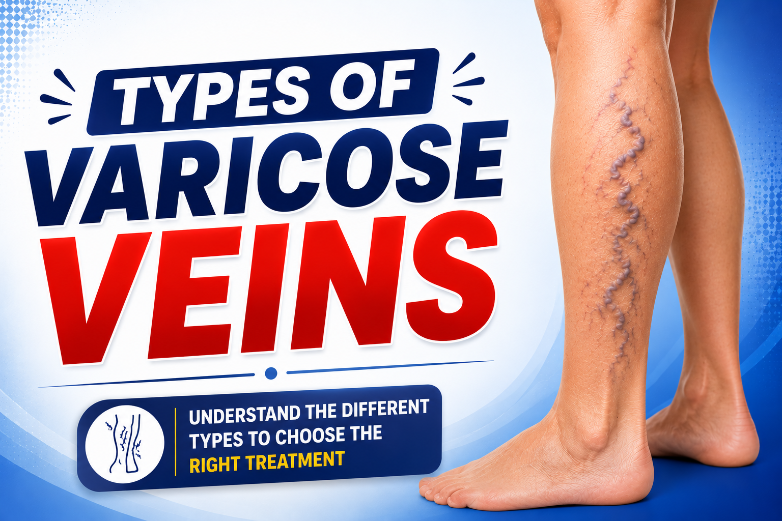
Truncal varicose veins are what most people picture when they think of varicose veins – large, bulging, twisted, rope-like veins that protrude visibly above the skin surface. They are 3 mm or larger in diameter and occur in the main superficial veins of the leg.
The two most commonly affected veins are:
Great Saphenous Vein (GSV) – runs along the inner side of the leg from the ankle all the way up to the groin. This is the most commonly affected vein in varicose vein disease.
Small Saphenous Vein (SSV) – runs along the back of the calf from the ankle to the back of the knee.
Where Do They Appear?
Along the inner leg, back of the calf, and behind the knee – following the path of the saphenous veins.
Symptoms
- Visibly bulging, twisted, dark blue or purple veins
- Persistent leg heaviness, aching, and fatigue
- Swelling around the ankles and feet
- Muscle cramps, especially at night
- Itching or burning sensation over the veins
- Skin discoloration in advanced cases
- Risk of venous ulcers if left untreated
Causes
- Failure of the one-way valves inside the saphenous veins
- Genetic predisposition – runs in families
- Prolonged standing or sitting occupations
- Pregnancy
- Obesity and sedentary lifestyle
- Age-related weakening of vein walls
Are They Dangerous?
Yes – truncal varicose veins are the most medically significant type. If left untreated, they can lead to:
- Chronic venous insufficiency
- Skin changes and discoloration
- Venous eczema
- Superficial thrombophlebitis (clotting and inflammation)
- Deep Vein Thrombosis (DVT)
- Venous leg ulcers – open wounds that are very difficult to heal
Treatment
- EVLT – Endovenous Laser Treatment – the gold standard treatment. A laser fiber is inserted into the diseased vein under ultrasound guidance and laser energy is used to seal it permanently. Minimally invasive, day-care procedure with 2–3 day recovery.
- Radiofrequency Ablation (RFA) – uses radiofrequency energy instead of laser to close the vein. Similar outcomes to EVLT.
- Foam Sclerotherapy – used for smaller truncal varicosities or as a complementary treatment alongside laser.
- Ambulatory Phlebectomy – small hook-like instruments remove varicose vein branches through tiny punctures. Often combined with EVLT for complete treatment.
Type 4: Perforator Vein Incompetence
What Are They?
Perforator veins are connecting veins that link the superficial veins (like the saphenous veins) to the deep veins inside the leg. Normally, they carry blood in one direction – from superficial to deep. When their valves fail, blood flows backward, increasing pressure in the superficial veins and worsening varicose vein disease.
Where Do They Appear?
Perforator vein incompetence does not produce visible veins on its own but significantly worsens existing varicose veins – especially around the ankle and lower leg.
Symptoms
- Worsening leg swelling and skin changes despite treatment of superficial veins
- Skin discoloration, hardening, and varicose eczema around the ankle
- Slow-healing or recurring venous ulcers
Are They Dangerous?
Perforator vein incompetence is strongly associated with advanced chronic venous disease – particularly venous leg ulcers. It must be identified and treated to achieve lasting results.
Treatment
- EVLT or RFA – used to ablate incompetent perforator veins under ultrasound guidance
- SEPS (Subfascial Endoscopic Perforator Surgery) – a minimally invasive surgical approach used in complex cases
- Treating perforator incompetence is essential for complete resolution of advanced varicose vein disease
Type 5: Pelvic Varicose Veins (Pelvic Congestion Syndrome)
What Are They?
Pelvic varicose veins are dilated, poorly functioning veins located within the pelvis and lower abdomen – rather than in the legs. They are caused by the same mechanism as leg varicose veins – valve failure and blood pooling – but occur in the ovarian and pelvic veins.
This condition is known as Pelvic Congestion Syndrome (PCS) and is a significant but frequently underdiagnosed cause of chronic pelvic pain in women.
Who Gets Pelvic Varicose Veins?
Pelvic varicose veins predominantly affect women – especially those who have had multiple pregnancies. Pregnancy increases blood flow to the pelvis dramatically, and in some women, this causes permanent valve damage in the pelvic veins.
Symptoms
- Chronic, dull, aching pain in the lower abdomen or pelvis
- Pain that worsens after prolonged standing, during or after intercourse, or during menstruation
- Varicose veins appearing in unusual locations – inner thighs, buttocks, vulva, or back of the knees
- Feeling of heaviness or fullness in the pelvis
- Occasionally associated with irritable bladder symptoms
Are They Dangerous?
Pelvic varicose veins are not life-threatening but significantly impact quality of life. Chronic pelvic pain is often misdiagnosed as endometriosis or gynecological conditions for years before pelvic congestion syndrome is identified.
Treatment
- Ovarian vein embolization – a minimally invasive interventional radiology procedure where a catheter is used to block the refluxing pelvic veins using coils or sclerosing agents
- Laparoscopic surgery – in selected cases
- Hormonal therapy may help control symptoms in some patients
Type 6: Vulvar Varicose Veins
What Are They?
Vulvar varicose veins are varicose veins that develop in the vulva (external female genitalia). They are closely related to pelvic varicose veins and often occur together.
Who Gets Them?
Almost exclusively occurring in pregnant women – particularly in the second and third trimester – due to the dramatic increase in pelvic blood volume and pressure from the growing uterus.
Symptoms
- Visible, bulging veins in the vulvar area
- Sensation of heaviness, pressure, or aching in the pelvic region
- Discomfort that worsens with prolonged standing or walking
- Occasional swelling in the inner thighs
Are They Dangerous?
In most cases, vulvar varicose veins improve significantly or disappear on their own within 6 weeks after delivery. However, a proportion of women retain them permanently and require treatment after pregnancy.
Treatment
- Conservative management during pregnancy – compression garments, rest, elevation
- Sclerotherapy or embolization – after delivery if veins persist
Type 7: Esophageal Varices
What Are They?
Esophageal varices are varicose veins that develop in the lower part of the esophagus (food pipe). Unlike other types of varicose veins, these are caused not by valve failure but by portal hypertension – increased blood pressure in the portal vein system, most commonly due to liver cirrhosis.
Symptoms
- Often no symptoms until the veins rupture
- Sudden vomiting of blood (hematemesis) – a medical emergency
- Black, tarry stools
- Signs of liver disease – jaundice, abdominal swelling
Are They Dangerous?
Yes – esophageal varices are a serious, potentially life-threatening condition. Rupture of esophageal varices causes severe internal bleeding and requires emergency medical treatment.
Treatment
- Endoscopic band ligation – a scope is passed into the esophagus and rubber bands are placed around the varices to cut off their blood supply
- Sclerotherapy – injection of sclerosing agent via endoscope
- TIPS (Transjugular Intrahepatic Portosystemic Shunt) – a procedure to reduce portal vein pressure
- Medications to reduce portal hypertension
Quick Comparison: All Types of Varicose Veins at a Glance
Type | Size | Location | Risk Level | Best Treatment |
Spider Veins | Very small | Legs, face | Low | Sclerotherapy, Surface Laser |
Reticular Veins | Small | Back of knee, thighs | Low–Moderate | Microsclerotherapy |
Truncal Varicose Veins | Large | Inner leg, calf | Moderate–High | EVLT Laser Treatment |
Perforator Vein Incompetence | Variable | Ankle, lower leg | High | EVLT, RFA |
Pelvic Varicose Veins | Variable | Pelvis, abdomen | Moderate | Embolization |
Vulvar Varicose Veins | Variable | Vulva, thighs | Low–Moderate | Compression, Sclerotherapy |
Esophageal Varices | Variable | Esophagus | Very High | Endoscopic Banding |
How Are Varicose Veins Diagnosed at Hari Laser Clinics?
Accurate diagnosis is the foundation of effective varicose vein treatment. At Hari Laser Clinics, every patient undergoes:
Clinical Examination – Your vascular surgeon carefully examines your legs in standing position to assess the distribution, size, and extent of varicose veins.
Venous Doppler Ultrasound – This is the gold standard diagnostic tool. It maps your entire vein network, identifies which veins are diseased, locates faulty valves, and detects any perforator incompetence or deep vein involvement. Without a Doppler scan, accurate treatment planning is not possible.
Based on these findings, your surgeon will recommend the most appropriate treatment – whether that is laser ablation, sclerotherapy, or a combination approach.
Frequently Asked Questions
Yes, absolutely. Many patients present with a combination – for example, large truncal varicose veins alongside spider veins and reticular veins. A Doppler ultrasound helps map out all affected veins so a comprehensive treatment plan can be created.
Not always. Spider veins can remain stable for years, but they can also indicate underlying venous insufficiency that may progress. A vascular assessment can tell you whether treatment is advisable.
Yes. Hari Laser Clinics offers the full range of varicose vein treatments including EVLT laser treatment, sclerotherapy, microsclerotherapy, and foam sclerotherapy – covering all types and grades of varicose veins.
Truncal varicose veins involving the great saphenous vein carry the highest risk of serious complications such as DVT, venous ulcers, and chronic venous insufficiency if left untreated. Esophageal varices are the most medically dangerous overall but are a separate condition caused by liver disease.
With modern laser treatment and sclerotherapy, the treated veins are permanently closed and absorbed by the body. However, new varicose veins can develop in other veins over time – particularly if underlying risk factors like prolonged standing, obesity, or hormonal changes are not addressed.
Conclusion
Varicose veins are not a single condition – they are a spectrum of venous disorders ranging from harmless cosmetic spider veins to serious truncal varicosities that can lead to dangerous complications if ignored. Knowing which type you have – and understanding its specific risks – is essential for making informed decisions about your treatment.
Whether your concern is cosmetic or medical, mild or advanced, early or progressed – there is an effective, minimally invasive solution available today. The key is to get assessed early, before the condition worsens.
Concerned about varicose veins? Get a complete vascular assessment including Doppler ultrasound at Hari Laser Clinics, Bangalore.